
Posted By:
In the day to day life of a vet in practice, one thing we often notice is just how ‘in tune’ owners can be with their pets, and how familiar they become with their usual habits and routine. So when an owner feels that something isn’t quite right with their pet, even if nothing specific is immediately apparent, we know they will usually be right. A good example of this is Chester the 11 year old Springer Spaniel and his owner.
Chester was brought in because his owner was concerned that he seemed a little subdued. They had also noted that Chester’s urine was perhaps a shade darker than usual and so they had sensibly brought in a urine sample. On first examination Chester appeared very well, and nothing abnormal could be found. His urine wasn’t drastically abnormal but we felt a quick test would help us rule out any potential abnormalities.
The dipstick test on the urine revealed red blood cells which shouldn’t have been there. With this finding we felt that more extensive blood and urine testing was warranted and so we sent the samples to the lab that day. The results were generally encouraging. Chester’s blood test was all normal and, although his urine analysis revealed signs of a heavy infection, at least this gave us a diagnosis – we could assume that Chester had picked up a urine infection and so started him on antibiotics, agreeing to monitor his progress.
Chester’s owner monitored him closely and completed his course of antibiotics. However, after an initial improvement he felt that Chester deteriorated and so brought him back to see us. Once again Chester appeared very well on first check. However, we were confident that his owner knew him well and that something wasn’t right, so decided to conduct more tests. Once again, these showed that Chester had a heavy urinary infection, something that really should have resolved with his previous treatment.
As we had used an antibiotic that we knew should effectively treat this infection we could find no obvious reason why Chester had not responded to his course of medication. This was an unusual situation so we had to investigate whether there was an underlying problem causing Chester’s condition.
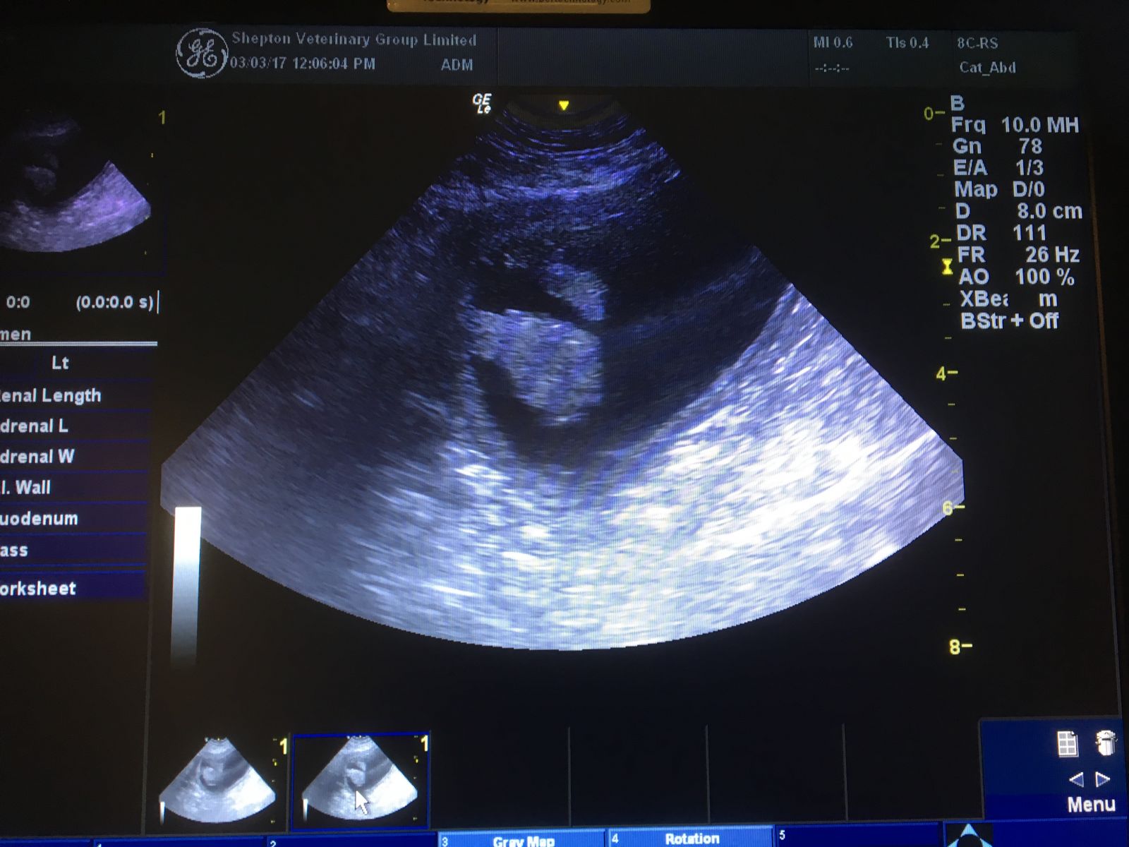
Chester visited for further testing and had an ultrasound scan of his bladder and x-rays taken. The x-rays showed no abnormalities but the ultrasound began to make things clearer. We were able to identify two abnormal growths at the front of his bladder. With Chester under general anaesthetic we could then pass a urinary catheter into his bladder and, using the ultrasound scan to visualise it, guide it on to the lumps. We could then apply suction to the catheter to enable us to collect a tiny sample of cells, which might help us establish what these growths were.
The lab test report on the cells taken from Chester’s bladder unfortunately advised us that these were tumour cells and that we should attempt to remove the lumps if at all possible. This was not good news and, naturally, we
need to discuss this at length with Chester’s owner. There was a good chance that we could remove the lumps completely and there would be no trace of any tumour cells left in Chester’s body. However, surgery to the bladder can be tricky, it may turn out impossible to remove the lumps completely and there was the possibility that tumour cells could have already spread through the body.
With Chester an otherwise very happy, healthy dog, his owner wanted to give him the best possible chance of a complete cure and long term recover. He had no hesitation in arranging the surgery.
Chester’s underwent an extensive surgery and in total 40% of his bladder was removed with the remaining bladder reconstructed. He was hospitalised on intravenous fluids until he had been seen to urinate and then he was discharged home where he was happiest whilst we awaited the results from the analysis of his removed bladder lumps. We continued antibiotic treatment for the infection and after a couple of weeks conducted urine testing again to confirm that his infection had resolved.
The biopsy results returned and confirmed that the two masses were indeed tumours and, although they had been completely removed, they were of a type that had the potential to spread. The lumps were also identified as the site of growth of the bacteria which had caused that persistent infection. The good news was that the infection had resolved completely and the removal of the lumps was definitely a huge positive, increasing Chester’s life expectancy. His owner also reported that Chester was back to his normal self.
After consulting with a veterinary oncologist, we have now started Chester on a special medication which has been shown to slow or prevent any recurrence of these tumour types. The main thing is that Chester seems to be back to feeling really well and we know that his owner will spot any changes, no matter how small, straight away.

Whilst on my visits I have been having several discussions...

As our feline friends get older there are a few conditions...

Another winter discussion group season is now behind...
©2024 Shepton Veterinary Group Ltd., All rights reserved.
Privacy Policy • Terms & Conditions • Cookie Policy