
Posted By:
Rosie is a lovely tortoiseshell female cat (with attitude !). Her owner noticed that she was having difficulty eating and one of her lower canine teeth was at an abnormal angle. On examination it was clear that the tooth was broken and the loose part was removed. The root was still present and, as retained roots can cause pain and infection, Rosie was booked in so that it could be x-rayed to assess whether it needed to be removed.
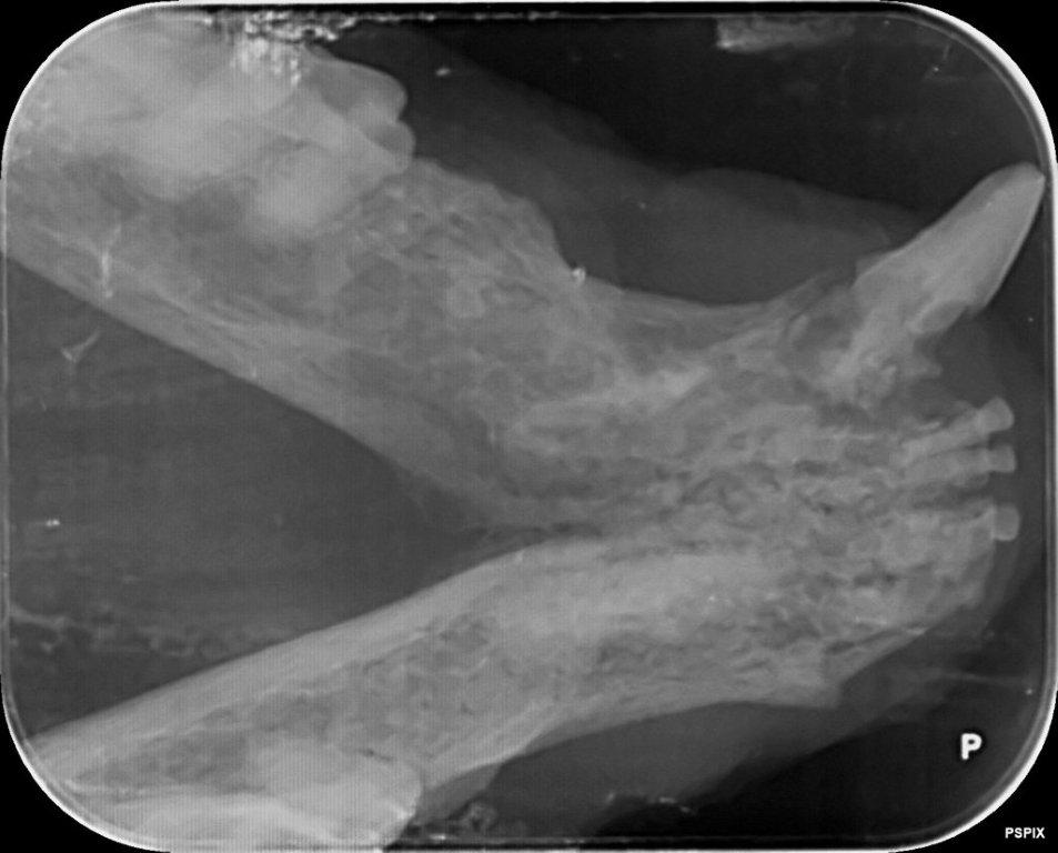
Under anaesthesia an x-ray was taken of her lower canines which clearly showed the remaining root. It also showed that the other lower canine had a significant lesion below the gum line due to ‘Feline Tooth Resorption’ (FTR). The x-ray attached is from a cat with similar issues to Rosie and shows the remaining root and the lesion below the gum line on the other side.
FTR occurs in cats where a lesion forms in the tooth around the gum line which is very painful. The lesion can progress until the tooth spontaneously fractures. It is not known why these lesions develop. It can be difficult to properly assess these lesions in conscious cats because they often sit just below the gum line and can only be identified by probing under anaesthesia or, where appropriate, by using dental x-rays.
We know that these lesions will progress and, along with the fact that they are painful, this makes removal of affected teeth important. There are 2 main types of FTR lesion and, in some, the root will be reabsorbed by the body. However certain roots do need removing and x-rays allow us to decide which ones this involves. Both roots were not seen to be resorbing in Rosie’s case so required surgical removal.
Removing lower canine teeth in cats can be quite difficult because the root is longer than the exposed crown and extends a long way in to the jaw bone. Lower jaws in older cats, especially if they have dental disease, can have quite poor bone density and they can fracture relatively easily when canine teeth are removed, particularly if both canines are affected.
Accessing the roots safely involves using a dental drill to remove the bone on the outside edge of the root to allow direct access to it and subsequent removal. The gum has to be incised first to access the bone and it is then sutured back together after the root has been removed. Using this method both of Rosie’s lower canine teeth were removed.
Because this surgery is quite invasive it can be quite painful in recovery so we ensured that Rosie had good pain relief to minimise any discomfort. After an initial slow recovery Rosie quickly felt better and, within 2 weeks, was completely back to her normal self.
Dental disease in animals can easily be underestimated in terms of the amount of pain it causes. It is often assumed that, if an animal is eating, their mouth is not that painful. However we know that animals feel similar levels of pain to us and many of us will know how debilitating dental pain is !
Together with the increased level of bacteria present with dental disease which can cause infections elsewhere in the body, this makes it important to monitor and recognise dental disease and to take appropriate steps to treat it correctly. This often means removal of affected teeth. Remember that an animal will be far happier and healthier with diseased teeth being removed rather than having a chronically painful mouth.

Whilst on my visits I have been having several discussions...

As our feline friends get older there are a few conditions...

Another winter discussion group season is now behind...
©2024 Shepton Veterinary Group Ltd., All rights reserved.
Privacy Policy • Terms & Conditions • Cookie Policy