Posted By:
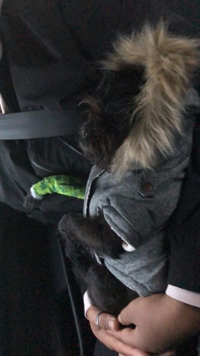
Pepsi is an 8 and a half year old female Pug who was seen by us as the last appointment during a busy Thursday evening surgery. She had been seen by another Veterinary practice near Yeovil where the owners would normally take her as she had developed gagging/retching type symptoms, especially after attempting to eat food.
She was initially treated for throat and stomach irritation/inflammation but after failing to improve on routine medications X-rays were performed to look for any potential blockage which might be occurring. Pepsi was known to love her food and had eaten some chicken bones the Sunday previously which may or may not have been significant.
The most common site for intestinal/gut blockages is towards the end of the small intestine as it starts to narrow, however they can occur at any site along the digestive tract. X-rays confirmed a blockage within Pepsi’s oesophagus (known as the food pipe or gullet) caused by a structure with a bony appearance which would need to be removed ASAP. Whilst the blockage remained in place Pepsi would be unable to eat or drink much at all, and the longer it was there the greater potential for irreversible oesophageal damage.
Pepsi was going to have to be referred for removal however the owners were friendly with one of our Farm Vets, Michael Head, who suggested they give us a call as we had only a few months ago purchased the equipment necessary for removal of such a blockage.
Pepsi was brought in and was soon placed under a general anaesthetic. The equipment needed for the removal is known as an endoscope. An endoscope is something commonly used in human medicine and is a long flexible tube with a camera at the end which can be passed down the digestive tract to allow visualisation of the oesophagus, stomach and small intestine. It can also be passed up the other end to visualise the colon, a so-called colonoscopy.
The endoscope was passed down Pepsi’s oesophagus and the bone obstruction was visualised. Another piece of equipment was passed alongside the scope which allowed us to grab hold of the bone and gently dislodge and retrieve it back up and out of the mouth. The oesophagus is quite a delicate structure and if damaged can be extremely difficult to fix given its location. If damage is severe it can cause a narrowing known as a stricture, which is due to a ring of scar tissue forming. Such a stricture can also block food passing down into the stomach.
Once retrieved Pepsi was woken up and given a few different medications to sooth the damage left behind and make her feel more comfortable. Fortunately it has now been 2 months since the procedure and she is doing extremely well. Any stricture would have formed by now but as she is eating fine and happy in herself we can presume that no significant stricture has formed and the oesophagus is back to functioning normally. Pepsi has made a full recovery, but her case does highlight the risks of feeding some bones to animals. Whilst they may have a place, caution must also be advised if feeding moderate sized bones which may be swallowed whole as these are the ones where blockages are most likely.
As it has turned out, the new Endoscope has so far been extremely useful in a number of cases similar to Pepsi’s (you may recall a recent social media post about the removal of a kebab stick swallowed by another dog!). It is also used for taking gut biopsies to diagnose conditions such as inflammatory bowel disease and I am sure it will continue to be of great use to us, yourselves and your pets.
Whilst on my visits I have been having several discussions...
As our feline friends get older there are a few conditions...
Another winter discussion group season is now behind...
©2024 Shepton Veterinary Group Ltd., All rights reserved.
Privacy Policy • Terms & Conditions • Cookie Policy