A Case of Adult Pneumonia
Published on: Dec 15, 2020
A high yielding AYC dairy herd of 180 cows began to experience snotty noses and coughing amongst the adult milking herd. It was mid-November, and soon these symptoms also became apparent in the heifers and calves. At housing there had been a change in the TMR and the cow cake, and the cows had gone loose and lost condition. There was an obvious drop off in fertility as oestrus activity was considerably reduced.
We were called on 3 separate occasions to sick cows. Some responded to antibiotic and NSAID treatment, but others didn’t. The herd vaccinated against IBR, and three animals that were swabbed, confirmed wild virus was not circulating.
3 cows that were unresponsive died and the fourth to die was taken for a post-mortem to Langford Pathology Service. A severe pleuropneumonia was diagnosed on post-mortem caused by the bacteria Mannheimia haemolytica (See Figs. 1-3).
Commonly associated in calves, outbreaks of Mannheimia haemolytica have been seen in adult herds, too. The photos show how large parts of the cows’ lungs were severely damaged as a result of the infection. The bacteria identified were sensitive to all known antibiotics, but it is worth noting sometimes they can be resistant to Oxytetracycline (Engemycin and Alamycin).
The approach to adult pneumonia outbreaks is the same as that in calf outbreaks, and assessment of nutrition, hygiene, and housing (including stocking rates) was done. Most aspects were very good, but the inclement and variable weather in November coupled with the dietary upset appeared to have been a tipping point.
As the cows adapted to the new ration and the weather settled, so the number of cases reduced. However, 5 cows, 5 heifers and 4 calves died – proving to be an extremely difficult costly disease outbreak. The plan is to vaccinate the adult herd against Mannheimia haemolytica in the New Year.
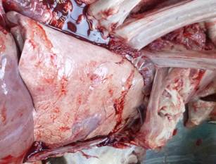
Fig. 1: A healthy cows’ lungs
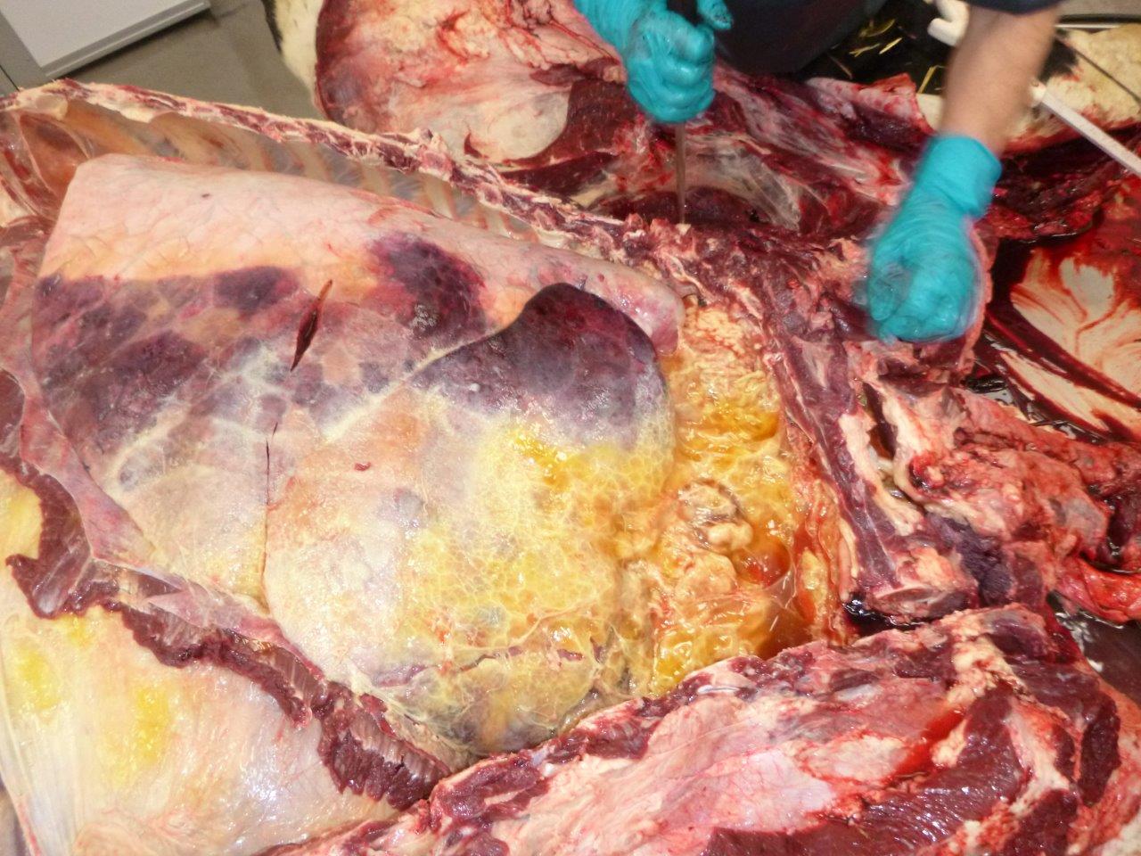
Fig. 2: Severe pleurisy – the right lobes with only a small area of normal lung remaining in the dorsal part of the caudal lobes
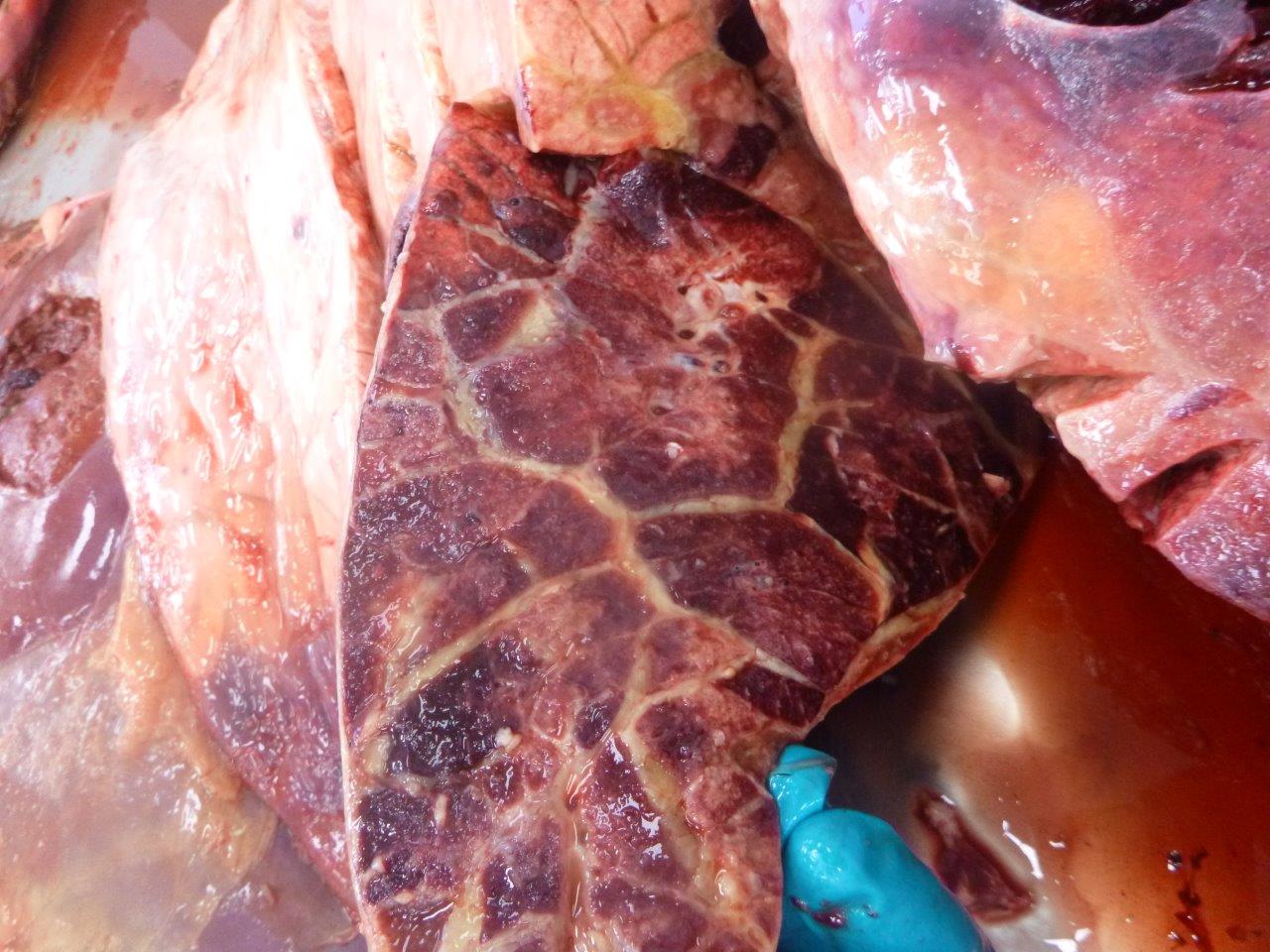
Fig. 3: Severe consolidation (no air spaces) and marked interlobular oedema (“marbling”)


