Pet Of The Year 2024
Rory
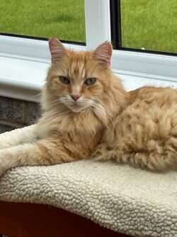
Senior feline patients can require some of our most regular and diligent care, from both owners and vets. Their age can predispose them to several medical conditions which can require regular vet visits, medications and specialised diets just to keep on top of their ideal bodyweight and general well-being. Three of the most common conditions in geriatric cats are chronic kidney disease, hyperthyroidism and diabetes – and Rory has been diagnosed over the last year with two of the big three!
Rory was 12 when he was first diagnosed with diabetes mellitus, after he was losing weight despite a ravenous appetite, developed an excessive thirst, and had started to flood the litter tray out with all too regular trips for a wee. As well as all of these symptoms, he had become very lethargic, more than just showing his older age. These are all the common hallmarks of feline diabetes, most similar to type II diabetes in humans. Diagnosed with a blood and urine test showing excessive sugar (glucose) in both, Rory ticked all of the diabetic boxes and was started on his treatment following an initial visit to the vets when his owner had become worried about him displaying the above traits.
Diabetes in cats is treated most usually by twice daily insulin injections administered by the owner at home, usually morning and evening around the time of a meal. Rory’s owner may have been initially daunted at the prospect of injecting him twice a day for the rest of his life but need not have been as she and him took to it effortlessly. Giving him insulin helped his body lock in his blood sugar, stopping him from losing weight and being so hungry and thirsty. It was slow progress at first as treatment had to start at a low dose before building up to find the right level of insulin for his body, guided by monthly blood tests. Alongside a specialised low carbohydrate diet for diabetic patients, Rory gained weight, and his symptoms improved.
Once settling on a dose of insulin, after he climbed back to his target bodyweight, all was not plain sailing as Rory then experienced a ‘hypoglycaemic’ episode where his blood sugar actually dropped too low and he started to tremor, feel weak, and even experience a couple of seizures. Following this, his insulin dose had to be readjusted, dropping right back to low levels before being slowly worked back up higher again. Rory’s blood sugar was becoming better controlled over a few months, but this time he unfortunately continued to lose weight.
After an additional blood test to check his thyroid levels, he was then diagnosed to be hyperthyroid – where his body was also producing too much thyroid hormone, causing him to have too high a metabolic rate, meaning that once again he was not able to gain weight. Another medicine was then introduced to control this second condition, this time in the form of a liquid on his food twice daily. Fortunately, once starting the medication, Rory gained weight again, and though he required further medication increases, he was once again returning to his normal self, no thanks to the dedication of his owner in persisting with injections, medications and frequent visits to the vet practice just to get him right again.
Bonnie

Old English Sheepdogs are a much loved breed in the UK – instantly recognisable for their long, shaggy coats and happy demeanour. Affectionately known as ‘Dulux dogs’ after their feature in a well-known paint advert in the past, they are less commonly seen these days. There are rescue charities dedicated to the breed, and Bonnie was rehomed to her current owners a few years ago as an older dog who had an unfortunate change in circumstances when her previous owners became deceased.
Over the past few years Bonnie has been enjoying a new lease of life in her ‘forever home’ that she shares with fellow senior rescue Old English Sheepdog, Bella. However, unfortunately neither are strangers to the veterinary practice and Bonnie was first seen in autumn of last year for a recurrent issue with her toe that just would not go away after she innocently stubbed it in the garden.
Initially Bonnie came back in from the garden with blood from her paw, but a few days later she started limping and had developed a swollen and smelly toe. Poor Bonnie was so lame she could barely put her foot to the floor and the second toe of her back right leg was covered in a discharge with a very foul odour indeed! Bonnie also had a temperature, so once her foot was cleaned up, she was advised to start pain relief and prescribed an antibiotic course to take with her meals for a nail bed infection. Unfortunately, at the end of the week her toe was no better at all. This being the case, she was admitted to have samples taken from her toe under sedation. There was no obvious cause or origin to the issue, but she was prescribed a different course of antibiotics and responded to these well over the following week.
The following month, Bonnie returned to the practice as her toe had swollen up again and started discharging bloody fluid. Antibiotics were restarted and Bonnie’s toe was X-rayed under sedation to rule out sinister causes or more serious trauma. The last little bone in her toe appeared to be partially dislocated and as it was connected to the infected nail, the decision was taken to remove the very end of her toe. Bonnie responded well to this minor surgery and further medical treatment and was able to enjoy her Christmas at home.
Just after Easter, Bonnie knocked her toe jumping in the garden and then became increasingly lamer and unwell. Another visit to the practice showed she had developed a fever, and a concern was she was becoming septic from a recurrent toe infection. Antibiotics again came to her rescue and Bonnie bounced back well, however at the start of summer her toe was bleeding more frequently and starting to really smell. Bonnie’s owners had been trying to manage the situation at home as best as could do, with fantastically designed homemade dressings, but eventually, we could not get away from the fact that her toe was causing too many problems, would now never fully heal and ultimately needed removal via amputation under general anaesthetic.
This procedure was not taken lightly as Bonnie was well over 13 years old now and had a heart murmur, but her owners felt she was otherwise well enough and that her quality of life could really benefit from this operation. The surgery went well, and with a few post-operative dressings changes her toe had healed well. Bonnie was managing to weight bear very capably on her back right foot despite missing a toe. Now toe-less she can hopefully happily live out the rest of her many retirement days with her dedicated owners.
Piper

Blood in our pet’s bodies is similar to most other mammals and therefore ourselves. As well as red blood cells carrying oxygen, and white blood cells helping to fight infection, another important component of blood circulation are platelets. These small cells are involved in blood clotting, and so are crucial to stop animals bleeding. If platelet numbers are low in our pets, this leaves them vulnerable to potentially life threatening haemorrhage. Piper the 2 year old cocker spaniel was found to suffer from low platelets when he appeared painful, and needed a 9 month course of medication to get his platelet numbers back up to scratch.
Dog’s platelets can be commonly reduced in number as a result of autoimmune disease, where they are destroyed by the body’s own immune system attacking them. This can result in a lot of minor bleeds throughout the body, shown as bleeding from any organ, in joint spaces, from the mouth and between muscles, which can result in pain, fever and lethargy – exactly the symptoms Piper had shown when he came into the vet practice having been off colour for a few days. Most commonly pain in muscles and legs of our dogs is due to strains, but when Piper wouldn’t respond to pain killers, and was later found to have a temperature and small bleed in his eye and gum, a blood sample was taken to investigate.
Suspicions were raised of immune mediated, or autoimmune disease, as young Cocker spaniels can be quite prone to these conditions. Later that day, the blood test revealed a very low platelet count in Piper’s circulation – in fact there were barely any seen in his sample! It was determined that Piper’s platelets had been attacked by his own immune system, so he would need medication to suppress his immune system, to allow his platelet numbers to build up again.
Steroids are the most commonly used immunosuppressive medications for our pets, and are usually given in tablet form at home. So Piper’s owner started a high dose course of daily tablets in the hope that his symptoms would subside. Unfortunately, as good as steroids can be, they can bring with them a lot of side effects, such as excessive hunger, thirst, urination and panting which Piper began to demonstrate. When Piper returned to the practice for a monitoring blood test, unfortunately it was found that his platelet numbers hadn’t really increased, so he needed an additional immunosuppressant to control his condition better. This was a drug known as mycophenolate, which can be quite powerful and show side effects of diarrhoea.
However, both of the medications alongside each other slowly started to work, Piper’s bloodshot eye resolved whilst he became brighter and more comfortable, and on each weekly visit to the practice, Piper’s platelet count would slowly rise. Along the way due to his body being immunosuppressed by the tablets Piper also picked up a tummy bug, but finally his normal platelet count was reached. It was at this point that Piper’s medications could be lowered. Immunosuppressants, especially steroids, need to be tapered or withdrawn very slowly to be safe, and a blood sample checked after each medication drop to avoid a relapse. 9 months down the line and Piper is now medication free having had his last blood test confirm a normal platelet count.
Milo
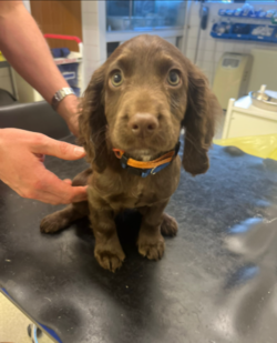
There are many surgeries and procedures performed at both our Shepton and Wells practice’s that we do routinely; neutering, lump removals, x-rays, scans etc… But every so often an opportunity comes along to perform a much rarer surgery, which in some cases we may never have done before.
Being presented with a litter of puppies or kittens often leads to us performing health checks, microchipping and vaccinations. In most cases no significant issues are found, perhaps a minor umbilical hernia now and again but often nothing that’s too concerning. Even heart murmurs in puppies and kittens are very common and if these are quiet, they frequently disappear by 6 months of age. As a rule, if a young puppy has a louder heart murmur, or they reach 6 months of age and a heart murmur is still present, this should then be investigated.
Milo came in to us initially as one of a litter of 6 puppies when he was only a few days old. He was brought in a few weeks later, along with all his brother and sisters as the owners could hear a heart murmur in one of his littermates, something they’d had confirmed by their neighbour who was also a vet. At this presentation Milo’s littermate was found to have a severe heart murmur (Grade 6/6) and Milo himself had a significant murmur graded 4/6 (the higher the grade the louder the murmur). Given the location, grades and sound of these murmurs it was highly likely that both puppies had a condition called ‘Patent Ductus Arteriosus’, known as a PDA. A PDA is where there is an open vessel connecting the two major blood vessels that leave the heart. This connection is meant to close at birth but occasionally it remains open. A PDA has serious consequences for the animal and sadly if left untreated animals rarely live past 1 year of age. A PDA is confirmed via an ultrasound heart scan but given the puppies size this would have been very tricky at this point. As any surgery wouldn’t be performed until they were bigger anyway, we opted to book both puppies in for an ultrasound scan at 5 weeks of age. Unfortunately, only 4 days later Milo’s littermate started to show signs of heart failure and had to be euthanised, likely due to a more severe PDA than Milo. Heart ultrasounds are considered tricky, and many practices will refer puppies for heart scans elsewhere or get someone in to perform the scan. Given my area of interest is Cardiology in particular, I was able to perform Milo’s heart ultrasound and confirm a PDA which was already causing some abnormal heart enlargement, but the heart was still contracting well.
The good news is PDAs can be treated and cured. There are 2 ways to do this. Firstly, the connection between the 2 vessels can be tied off during open chest surgery. Secondly. more recent developments have allowed vets to perform a less invasive treatment whereby a vessel occluder is threaded up an artery in the leg to the heart and positioned in the connection between the vessels to block the blood flow. This latter procedure requires moving x-rays (fluoroscopy) and is only performed at referral centres. It also carries a significantly higher cost. As puppies with heart conditions are often not covered by insurance affordability of treatments can be a big factor. We had never performed a PDA surgery before but have done multiple open chest surgeries and offered to attempt the operation on Milo as his only affordable option of a cure. The owners were aware of the surgical risks and wanted to proceed.
Milo was operated on at only 10 weeks of age. He had a repeat ultrasound on the day of his surgery and the surgery itself involved 4 members of the team, 2 vets and 2 nurses. A meeting was held before the surgery to discuss the anaesthetic considerations and possible complications, and how we might address these if they were to arise. His surgery went well and the connection between the vessels was tied off successfully. On recovery Milo ‘s heart murmur was gone, and he was up and eating that same evening. Puppies are extremely resilient, and he was able to go home the next day having been monitored throughout the night. All that’s left is one final heart scan to in 2-3 months’ time to confirm that the abnormal heart enlargement has reversed, and Milo should ultimately now lead a normal life.
Only by having such a diverse range of interest and skills were we able to offer this previously considered ‘referral-only surgery’ at a cost that meant we could cure Milo’s condition. Various members of our staff were involved throughout the process, from ultrasound scanning to confirm the diagnosis, performing the surgery itself, monitoring the anaesthetic, and providing 24-hour post-operative care. Having been able to provide this treatment for Milo the aim now is to offer the same procedure to any future puppies found to have the same condition so they can live a normal happy life.
Baby
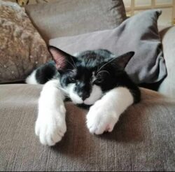
Our pets are a huge source of pleasure in our lives. Sometimes however, they can also be a source of stress and heartache. And this is definitely the case for Baby the 3 ½ year old cat, although I am sure his owners think he’s completely worth it.
Several years ago Baby used up one of his 9 lives when he was involved in a road traffic accident, and he presented to us shocked and with a fractured back leg. On that occasion he was stabilised, given pain relief and had surgery to repair the fracture. After several months of recovery, and a lot of hard work from his owner, he was back to himself. The main hope was that lessons were learned and he would come out of the experience more street wise.
Things had been great for Baby until one Friday afternoon when he started having difficult breathing. His owners knew this was not a time to just ‘wait and see’ and so contacted the surgery straight away. Within an hour Baby was examined by Vet Josh.
Josh could immediately see why Baby’s owners were concerned and so admitted him for hospitalisation in an oxygen chamber. The oxygen-enriched environment made life a bit easier for Baby and, after a few minutes, he started to look more relaxed, allowing some time to consider a plan to find out the cause of Baby’s difficulties.
The first step was an ultrasound scan of his chest as it is a non-invasive procedure that we could perform without stressing his breathing too much. Our main aim was to determine if there was any fluid in his chest, which is always an abnormal finding. However, once we started the scan we could see the problem was not fluid in his chest but something else – the image we saw resembled intestines, which definitely don’t belong in the chest. We started to understand what was happening, and after taking an x-ray the problem was confirmed – Baby had a diaphragmatic hernia.
A diaphragmatic hernia is a hole in the diaphragm – the thin muscle membrane that separates the chest and abdomen – which can allow the abdominal organs to move through the hole into the chest cavity. This causes multiple issues but most significantly can prevent proper expansion of the lungs and impact on breathing. It is usually the result of trauma, such as being hit by a car, in which the sudden increase in abdominal pressure causes a tear in the diaphragm, but the effects can take a while to develop as abdominal organs can move slowly into the chest over time. When we analysed Baby’s records we could see that he had lost a considerable amount of weight over the previous few months so it was clear that he must have suffered a trauma some time before but was only just starting to suffer the effects on his breathing.
With the diagnosis established we now needed a plan to treat him. Baby’s mild sedation and oxygenation meant his breathing had definitely improved and he looked more stable. We knew that the diaphragmatic hernia would require surgical repair but we also knew that we shouldn’t rush into this straight away. Both the anaesthesia and surgery is tricky for these patients and there is plenty of evidence that outcomes are better if it is delayed until patients are properly stabilised. So Baby was hospitalised and monitored overnight in the oxygen chamber, while we made sure we would have a full anaesthesia and surgical team, plus the required kit, ready for the following day.
Baby had a settled night and the next day, while some of the team were managing a busy Saturday of appointments, we proceeded with his surgery. Nurse Daisy was managing Baby’s anaesthetic, which she knew would be challenging – once we opened Baby’s abdomen to operate the negative pressure around the lungs would be lost and they would not expand normally. This meant that Daisy was going to have to manually inflate Baby’s lungs and breathe for him, carefully controlling his oxygen and carbon dioxide levels, whilst also controlling the depth of his anaesthesia and maintaining his blood pressure and fluid balance. Meanwhile Nurse Nat would ensure we had all equipment and medications needed during the procedure and Vet Gudi would assist me with the surgery.
As we started the surgery we could immediately see the extent of the problem, with much of Baby’s abdominal contents sitting within his chest. The surgery involves replacing all of the herniated organs back into the abdomen, while handling them delicately to avoid tearing or damage, and slowly we retrieved half his liver, as well as his pancreas, spleen, small intestine, right kidney and a section of large bowel back to where they belong.
After placing a surgical drain into his chest we stared to repair the hernia. It was in a difficult location to reach and we needed to place sutures through the torn muscle and around his ribs and body wall to close the defect. Once we were satisfied that our repair was good we finally started to close his abdomen whilst monitoring to see if Baby would start to breathe by himself.
Seeing that first breath after such major surgery is always immensely rewarding so we were over the moon when Baby very quickly started breathing on his own. Baby was closely monitored in recovery and we regularly checked his chest drain for fluid and air in his chest, which can accumulate during the surgery. This subsided very quickly and, with Baby settled and comfortable, we were able to remove his drain and send him home within 24 hours.
Baby has continued to do well, in no small part due to his owner’s diligent care. He’s clearly managed to get himself into a fair bit of trouble over the years, but has done so well through his two major operations because of his owner’s dedication to him. Hopefully now he really has learnt his lesson and might just let his owners enjoy their time with their beloved pet!
Thatcher
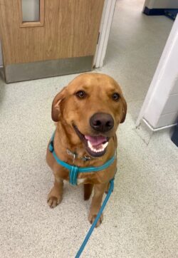
When Thatcher was brought into the surgery a month ago, it was clear that something was seriously wrong. He had been seen the day before with some non-specific abdominal tenderness but had deteriorated by the next morning to the point where he had to be carried in by his worried owner to see Vet Sarah.
On a physical examination, it was a bit of a mystery as to why he was so poorly and had such a painful tummy, particularly as he wasn’t vomiting and had no diarrhoea. He was a little pale and had a high heart rate – the latter was thought to be associated with the severe level of abdominal pain that he was experiencing.
Thatcher was admitted for hospitalisation and immediately started on strong pain relief to make him more comfortable as well as intravenous fluids.
A blood sample was taken to try and work out what was going on. Half an hour later, nurse Daisy who was caring for him noticed some blood around the site of his sample, and when we checked, we could see some bleeding from the site. This is very unusual and suggested a problem with blood clotting. We are fortunate to be able to rapidly test clotting times ‘in house’ so a fresh sample was taken from his leg which could then have a pressure bandage applied to stop further bleeding. The result from the test was not a normal or prolonged clotting time but ‘no clot’.
There may be several different causes of clotting disorders in dogs, but looking at the rest of his blood tests, it was a pattern that is typical of rodenticide poisoning. Abdominal ultrasound and chest and abdominal X-rays were checked to ensure no other explanation could be found. We are aware that rodenticides can cause abdominal pain, usually associated with bleeding into the gut.
At this point, it was essential to get clotting factors into Thatcher as he was in a critical condition. Vet Rachel had taken over his care at this point and suggested that we gave him a transfusion of fresh frozen plasma that we had stored at the practice. The plasma will contain active clotting factors so is very useful in this type of case. Thatcher was blood-typed first in case he needed multiple transfusions, and then the appropriate plasma could be used. He was also started on high doses of Vitamin K which is the antidote to anticoagulant rat baits.
The next day, he was a lot brighter and had started to eat, which is always a good sign for a labrador. His blood count had dropped, we felt this was likely due to bleeding before the treatment had taken effect but kept him in again overnight as a precaution.
When I went in to see him the following morning, he was like a different dog, the sad eyes were gone, and replaced by a dog who was clearly looking for his third breakfast (and the nurses can never resist those pleading eyes). His clotting times and blood count were back up to normal and he was able to go home.
Thatcher had to stay on Vitamin K for 3 weeks and has had his clotting times checked twice.
It is a mystery where the bait came from. We knew that small amounts of a bait with low toxicity for dogs had been in the area but can’t account for something so serious. Rat baits vary hugely in how much a dog needs to consume before toxic effects are seen, and the effect can be cumulative over time, so it’s possible that Thatcher had come across a concentrated bait at some stage and ate some without anyone seeing him.
We’re just glad that he’s back on good form and enjoying life at home with his brother and family.
Primrose

This is the story of sweet little Primrose, a 1-year-old cockerpoo. Primrose first came to see me back in April as her owners had noticed that she was having some blood in her wee. She was her usual happy self otherwise and eating and drinking all okay. On examination there was nothing that I was too worried about so it was most likely that she had picked up a urinary tract infection so we started her on some antibiotics and pain relief in the hope that this would clear things up. Unfortunately, this was not the case, and I resaw Primrose a couple of weeks later with the same issue. We decided that it might be best to send a urine sample away to the lab just in case it was a more complicated infection and make sure there were no crystals or stones causing the issue.
This is where things became interesting. The urine sample came back showing that there was indeed an infection present but there were also ammonium urate crystals present in Primrose’s wee. Now, these crystals are not often seen in the urine of healthy animals. They are more commonly found in dalmatians, which Primrose clearly was not, and can be an indicator that there is an issue with the liver. The livers’ role in the body is to clean the blood and remove build-up of toxins such as ammonia. If this is not working properly then these toxins must be excreted another way, for example through the kidneys in urine.
In a dog as young as Primrose, this made me concerned about something called a portosystemic shunt. Before a puppy is born, and is in the uterus of the mother, the puppies’ blood is cleaned by the placenta rather than the liver. Blood is diverted around the liver by a connection between the portal vein and another blood vessel. Usually when a puppy is born, this connection closes to allow blood to flow through the liver and be cleaned. In some cases, this connection does not close properly causing a shunt. This can occur outside the liver (extrahepatic) or inside the liver (intrahepatic). As a rule, small to medium dogs and cats have extrahepatic shunts, whereas larger dogs have intrahepatic shunts, though there are exceptions to this generalisation. Without treatment a shunt will cause a build-up of toxins in the body, potentially causing seizures and neurological issues and eventually death.
To investigate further we booked Primrose in for some blood tests and an ultrasound scan of her tummy. These showed changes in her blood consistent with a poorly functioning liver as well as elevated bile acids. She also had lots of small stones present in her bladder and her liver was abnormally small for a dog her age. All these things made a diagnosis of a liver shunt extremely likely.
Shunts can be treated one of two ways. Medical treatment involves a change of diet and lactulose to prevent build-up of toxins in the blood. Whilst this can be successful in the short term, surgical repair of the abnormal connection is the best treatment as this solves the issue and allows normal blood flow through the liver. Without surgery life expectancy is limited to 1-2 years maximum. This being said, surgery is very risky and close monitoring is needed post-surgery to ensure there are no complications. This means surgery can only be performed at specialist hospitals and so, because Primrose was otherwise well, a referral was made to Langford vets in Bristol for a few weeks’ time. Primrose was started on some medical management in the meantime to keep her stable until the time of surgery.
At Langford Primrose had a CT scan which confirmed the shunt which was extrahepatic and then proceeded to surgery. A device was placed around the abnormal vessel to allow gradual closure and her bladder was flushed of any stones present. She spent 5 days in the intensive care unit after her surgery and was monitored closely whilst she recovered. She is now back at home and recovering well from surgery. Her wound is healing nicely, and she is still her bright happy bouncy self. She will have to continue with her special diet for now whilst her liver gets used to its new blood supply but will be having blood tests in the next few weeks and if they are all normal this can be stopped.
This just shows how sometimes something simple can turn out to be something more serious as well as things not always presenting as written in the textbooks. Because Primroses’ owners noticed there was something wrong early on this meant we were able to treat her before she became unwell and can now go back to being her fun-loving bouncy self!
Loki
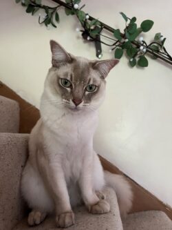
Oral health is a common area of concern in our dogs and cats, and we frequently see dental disease at the surgery. In the case of dental disease, it is often necessary to extract severely affected teeth and many older patients have to have multiple teeth removed to treat their condition. Loki the 4-year-old Burmilla cat had to have his teeth removed for a more unusual reason however, and at a much younger age than the average cat – which Loki most certainly is not!
Loki is a real character and extremely vocal. He and his brother life an indoor lifestyle with lots of environmental enrichment to provide for their needs and keep them happy. As we saw Loki through his first year of life, he was doing well with no major health concerns. However, during his first vaccination booster at 1 year old, we noticed that he had an area of gingivitis – inflammation of his gums.
Gingivitis is a common finding, usually developing as a reaction to a buildup of plaque, the invisible film of bacteria that accumulates on teeth. As such we usually see it in older patients as the plaque has built up over several years. However, Loki was very young to have this symptom and there seemed to be no problem at all with his teeth. As the gingivitis was not severe, at that stage we thought it might be a transient finding and possibly related to some stress at the time. We advised his owner to monitor it but discussed how to slow plaque build-up through toothbrushing and dental diets.
However, a year later when Vet Josh saw Loki for his next booster his gums were much worse. His owners had been taking steps to look after his dental health at home but had noticed the gingivitis continuing to develop and when Josh examined him it was clear that the inflammation was severe, particularly at the back of his mouth, and very uncomfortable.
Josh explained that Loki appeared to be suffering from Feline Chronic Gingivostomatitis, a complex disease in which the gums and other oral mucosa become severely inflamed, extending beyond the normal reaction to dental plaque. It’s a condition that we don’t fully understand yet but is considered to be caused by an inappropriate immune response to the normal oral bacteria, with possibly other triggers involved. And while there are a lot of possible helpful treatments suggested, the most effective option at the moment is to extract the patient’s teeth.
We explained that studies suggest at least two thirds of cats with CGS will have a complete or substantial improvement in their condition with extraction of only the teeth related to the inflammation, with some cats then requiring the remaining teeth extracted. Loki’s owners were understandably daunted by the prospect of him having any teeth extracted at his age, let alone potentially all of them. However, they could see the severity of inflammation and had noticed that it was causing him distress at home. After some thought and discussion, they booked him in for extraction of all his premolars and molars, which were the teeth closely associated with his inflammation.
Loki came in a few weeks later for his procedure. He was anaesthetised and had dental Xrays taken before we proceeded with the extractions. Extracting healthy teeth can be a difficult process, as the roots are strongly embedded in their sockets, and it is important that the roots are completely extracted. Loki’s gum tissue was surgically lifted away from the bone and we carefully and slowly extracted his back teeth, leaving all his canines and incisors in place, before suturing the gum closed and taking the post-extraction Xrays.
Loki had local anesthetic and painkillers during his procedure and was sent home with plenty of pain relief too, but he initially really struggled in his recovery. He didn’t want to eat for several days, he was unusually grumpy and his brother was avoiding him as well. With some diligent care from his owners and some extra medications he finally turned a corner. He seemed back to his normal self, happy and eating dry food, for a few months.
However, we were soon back to see us as he was suffering again. His owners had noticed that he would go through periods when his character would change – although it did not stop him eating, he would become withdrawn, agitated and more vocal than usual, and they could see that his gums were bright red again during these episodes.
His owners were worried about removing the rest of his teeth so we discussed other options that we could try first. We opted to trial a hypoallergenic diet which may reduce the antigen triggers in his mouth, alongside a combination of painkillers and antinflammatories to help relieve his symptoms. Then, when he developed gastritis with one of the medications we tried some alternatives, including an immune-modulating drug. Over the period of four months, we had the same repeating pattern – everything we tried seemed to help him for a while but then his mouth would get worse, and Loki’s poor owners had the heartbreak of seeing him withdraw and miserable again.
In the hope we could produce another solution we spoke to a feline medicine specialist for some advice. This was really helpful, and we came away with some tests we could run to rule out some complicating problems and some possible treatments which might help, but ultimately the advice confirmed what we already suspected, that Loki’s best chance of treatment was by extracting the rest of his teeth.
Loki came in for his second dental extraction procedure and had his remaining 16 teeth removed. Post-extraction Xrays confirmed there were no root fragments remaining and Loki was recovered from his anaesthetic. This time we were fully prepared for him to be a grumpy boy and, true to form, he took some time to recover. However, after a few checks at the surgery and his strong pain relief he quickly started to get back to his normal self.
I was delighted to receive an email from his owners two months later to let me know that he was like a new cat! They were amazed how well he had taken to his new toothless life, mouthing his toys and eating dry food quite happily. He had no further symptoms of his sore mouth and his temperament was much more settled. We have since seen him twice at the surgery and have been really pleased to observe that his gums looked healthy with no sign of the soreness he had before.
Extracting all of Loki’s teeth at just 2 years old was a brave step for his owners to consider and not one that we undertook lightly. It was through working together to discuss and research his options, and his owners taking the time to consider what was right for him, that we were able to find a solution for his painful condition – even if it does mean he is a bit gummy!


