A Closer Look: How Veterinary Endoscopy Helps Us Find and Remove Foreign Bodies
Just recently, we have had a bit of a run on retrieving foreign objects (medically referred to as foreign bodies) from both dogs and cats using our endoscopy equipment.
It has recently been ‘grass seed season’, and it is very common for these seeds to become embedded in paws, get stuck down ears and ultimately make their way through all different areas of the body. They can be very destructive and cause huge amounts of damage and inflammation as they migrate. Some of the most serious cases we see are often in spaniels that have inhaled them.
Alongside these seasonal foreign bodies, however, we still have to contend with all the others that can occur all year round. We know that dogs in particular like to eat things they shouldn’t, and these things can subsequently get stuck. For those that land and remain in the stomach, we try to avoid surgery where possible by using our endoscopes.
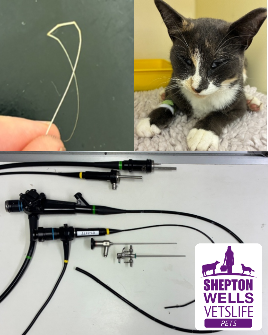
A veterinary endoscope is essentially the same as those used in human medicine, consisting of a flexible tube with a camera on the end that allows us to look inside the stomach and intestine. The endoscope attaches to a TV screen, giving us an enlarged image. The tube also has a small central channel through which we can pass instruments, allowing us to grab or ensnare objects and remove them back out through the mouth. This is often a lot fiddlier than it sounds!
In the last week, we have successfully done this and removed both a plastic disc and a Magnum ice cream stick. Keep an eye out on our Facebook and Instagram pages for the video of this removal, which will be uploaded on 10/10/26.
Whilst the endoscope used to retrieve objects from the stomach is quite large, we also have a smaller endoscope, known as a bronchoscope, which can be passed down the airways of animals. This allows us to look for abnormalities within the lungs or, again, to find and retrieve foreign bodies.
Most recently, we had a cat called Pickle in, who is just over two years old and had been coughing, wheezing and having episodes of shallow breathing for around a month. Given her age and clinical signs, asthma was considered the most likely cause. However, when she didn’t respond to medication, we opted to investigate further.
Pickle was anaesthetised and, after assessment of her larynx and vocal cords, a swab was taken from her trachea. The idea was that this swab could be sent away to look for specific viruses or bacteria. To my surprise, when I removed the swab, a strand of what was later thought to be one of her whiskers came out with it! Certainly enough to be irritating her airway and potentially causing her signs.
We followed this up with a couple of chest X-rays, which appeared normal, and used the bronchoscope to look down her airways and ensure that no further foreign bodies were present (they weren’t).
Pickle recovered from anaesthesia well and went home with her owner later that day with the plan to slowly reduce medication. During her post-op phone call, we were delighted to hear her owners report that she has recovered well and is back to her usual self – leaving us with the conclusion that the pesky whisker was the cause of the irritation all along. Thanks to the bronchoscope kit, we were able to thoroughly assess Pickle and confirm that there were no further issues, all without the procedure being too invasive.
The examples above demonstrate how we use our flexible scopes on a regular basis to remove objects, but these are also used to take biopsies, particularly in cases involving ongoing vomiting or diarrhoea.
Alongside our two flexible endoscopes, we have another, shorter, so-called rigid endoscope. This is the primary scope used to look up an animal’s nose (rhinoscopy) and gives the clearest picture of them all. It allows us not only to remove objects lodged up the nose, but also to investigate causes of ongoing nasal signs and take biopsies.
This rigid scope is also used to look within a female dog’s bladder (cystoscopy), allowing, amongst other things, the confirmation of any malformed ureters.
With the use of these scopes, we are able to visualise internal areas of the body that we would otherwise be unable to access without large open surgeries, obtain diagnostic biopsies with minimal discomfort to the animal, and retrieve those pesky foreign bodies that we see all year round in the airways, nose and stomach.
Author –