Posted By:
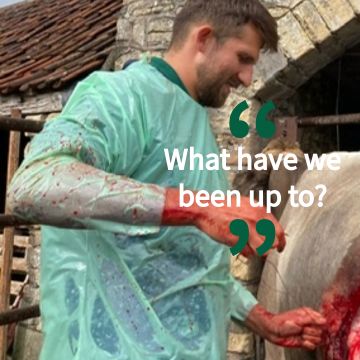
As the practice continues to grow and each vet develops their own areas of interest it is not unusual for cases to require input from more than one of us to achieve the best outcome. An example of this is Eddie, a 10-year-old Patterdale terrier. He initially came in some months ago due to back-end irritation and had been scooting along the floor and licking himself, something we commonly see due to anal gland problems. Eddie had these emptied a couple of times, but the irritation persisted, and I re-examined him 3 months after they were last emptied due to little improvement. At this consultation there was some skin ulceration in the region of the left anal gland which was now associated with a lump in the same area. This raised two main possibilities – either the lump came about because of the area being inflamed for a long period of time (option 1), exacerbated by Eddie’s licking, or it represented a genuine mass (option 2). It is impossible to tell such lesions apart by looking and so a sample was required. It’s a very sensitive area, so Eddie required an anesthetic to allow us to do this. Two types of samples were taken – a needle biopsy (due to its quick turnaround time) and a tissue biopsy which takes longer to get the results. The needle biopsy initially suggested an inflammatory lump (option 1) however the tissue biopsy showed a genuine mass/tumour (option 2) known as a carcinoma. In these situations, tissue biopsies are always superior and so this was the correct diagnosis.
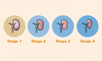
Now that we knew what we were dealing with we had to get it removed. The issue was that, like many other lumps, carcinomas associated with the anal glands can often spread to other areas. Such anal sac adenocarcinomas generally spread to the local lymph nodes (local glands) first before progressing elsewhere and so we wanted to see if these appeared normal. Some of these glands can be palpated rectally and some can only be visualized with imaging such as ultrasound. The rectal lymph nodes felt normal. The internal lymph nodes require the ultrasound to be seen but this Is not routinely performed and required some advanced ultrasound knowledge. Whilst I do a lot of imaging, especially ultrasound, I do not do as many complex surgeries. Eddie was therefore booked in on a day when I was available to image the internal glands and another vet, Martin, was available for the surgery, allowing us both to use our particular interests together. The glands/lymph nodes (known as the medial iliac lymph nodes) on the ultrasound appeared normal and measured within normal size limits. Whilst this can never be 100% accurate it allowed us to proceed with surgery with more confidence that it was the right decision. After removal the mass was sent for analysis and appeared completely removed in what is quite a tricky location. Eddie has recovered well and whilst there is still the potential for microscopic spread to have occurred pre-surgery, the best we can do now is monitor the surgical site for signs of recurrence. Different vets performing certain aspects of a workup like this is something we do very regularly and is likely to occur with more and more frequency as we each continue developing our skillsets in certain areas to provide better overall care for your pets.
- Vet Josh
Eddie's story underscores the importance of teamwork and specialization within our practice. As our veterinarians continue to hone their skills and expand their expertise, we remain committed to delivering comprehensive care that ensures the health and happiness of your furry family members. Thank you for entrusting us with their well-being.

Whilst on my visits I have been having several discussions...

As our feline friends get older there are a few conditions...
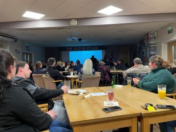
Another winter discussion group season is now behind...
©2024 Shepton Veterinary Group Ltd., All rights reserved.
Privacy Policy • Terms & Conditions • Cookie Policy